Treatment for Your Child's Hypoplastic Ventricle: Stage I
Stage I
The most common treatment for a child with heart problems that include a hypoplastic ventricle is heart surgery. Treatment is complex and requires careful management of your child’s health. It does not repair your child’s heart problem. But, it can relieve symptoms and increase your child’s chances to live a more normal life. This is often done in three stages. This sheet helps you understand the surgery that is done in the first stage of treatment.
The goals of heart surgery
- Make the single working ventricle the main pumping chamber of the heart. It will send oxygen-rich blood to the body.
- Decrease the workload of the single ventricle.
- Separate the circulation of blood in the heart
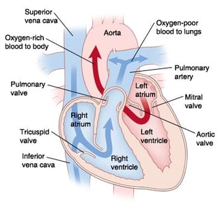
so that oxygen-poor blood and oxygen-rich blood don’t mix. - In a normal heart, oxygen-poor blood is pumped to the lungs from the right ventricle. Oxygen-rich blood is pumped to the body from the left ventricle.
Risks and Possible Complications of Heart Surgery
- Arrhythmia (abnormal heart rhythm)
- Problems with breathing
- Infection
- Bleeding
- Problems with the nervous system
- Too much fluid around the heart or lungs
- Difficulty feeding
Stage I: The Norwood Procedure
This procedure is generally done within the first week after birth. A hospital stay of 6 to 10 weeks may be needed. During the procedure, the surgeon does the following:
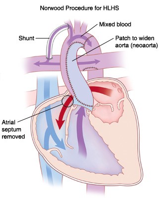
- Atrial septectomy. The wall dividing the two upper chambers, called theatrial septum, is removed. This lets oxygen-rich blood from the left atrium mix with oxygen-poor blood in the right atrium.
- Reconstruction of aorta. The main pulmonary artery is divided and used with patch material to remake the aorta. This is called the neoaorta or “new” aorta. Blood from the right ventricle can then be pumped through the pulmonary valve to the new aorta. This sends blood directly to the body instead of to the lungs.
- Placement of shunt (tube). A new path must be made to send blood to the lungs. This is because the main pulmonary artery has been used to remake the aorta. There are two different ways of making a shunt:
– Blalock-Taussig Shunt. A shunt is placed to connect an artery branching from the aorta to the pulmonary artery. This lets a controlled amount of blood reach the lungs (shown in picture).
– Sano Modification. A tube is placed from the right ventricle to send blood directly to the pulmonary artery (not shown).
Home Monitoring Program
Your baby will go home with a baby scale and a machine that measures the amount of oxygen in your baby’s blood. It is called a pulse oximeter. You will be given a book or application to download onto your smart phone. This will keep a record of your baby’s feeding schedule, oxygen level, and weight. The cardiology nurse practitioners will teach you how to do this before your baby leaves the hospital. They will let you know when and who to call if you have questions or concerns.


