Upper GI and Small Bowel X-Rays
An upper gastrointestinal with small bowel (UGI with small bowel) is a test that uses x-rays to look at the esophagus and the stomach. The esophagus is the tube that carries food and drink from the mouth to the stomach. The test also includes the first part of the intestine that carries the food out of the stomach. This test helps to see how well these organs are working.
Your child’s Upper GI X-ray is in Imaging (Radiology) Department at Children’s Wisconsin Hospital.
Please stop at a Welcome desk to get a badge and directions to Imaging.
To get a good test, the stomach and small bowel need to be empty before the x-ray pictures are taken. Follow the directions below based on your child’s age:
0 to 6 months old. Nothing to eat or drink 3 hours before the test.
7 months to 3 years. Nothing to eat or drink 4 hours before the test.
4 years and older. Nothing to eat or drink 6 hours before the test.
Important Information
- It is important that you follow these special instructions. If your child eats or drinks anything after the times listed above, the x-ray may be cancelled.
- If your child had x-rays or other images done somewhere besides Children’s Wisconsin, please bring copies of the images when your child has their test. This includes CT scans, MRI, Nuclear Medicine tests or Ultrasound.
- Please bring a list of all your child's medicines.
Before the x-ray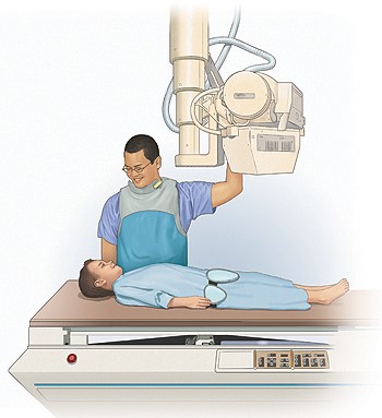
When you come to Imaging someone will greet you. Your child may be asked to put on a hospital gown and robe. A person skilled in taking X-rays, called a technologist will come and explain the test to you and your child. Please ask any questions you have. The technologist will take your child to the room where the test will be done.
How the test is done
- Your child will lie down on the x-ray table for part of the test. An x-ray will be taken before the test begins.
- During the test your child will drink thick, flavored liquid called barium.
- A large camera will take x-rays of your child as they drink the barium. The camera can see the barium inside the body.
- A doctor who is an expert in taking and reading x-rays will watch the picture on a TV screen. This doctor is called a radiologist.
- When the doctor is done taking x-rays, your child will go back to the waiting room. Your child may need to drink more barium.
- The technologist will take another x-ray about every 30 minutes. X-rays will be taken until the barium gets to the end of the small bowel (intestine). When that happens, the doctor will take one last picture.
Parents: If you wish to stay with your child during the exam you must meet the safety guidelines.
- You cannot have other children with you and you must not be pregnant.
- Parents who stay in the room must also wear a lead apron.
How long will the test take?
Each person is different, but this most often takes from 1 to 3 hours, sometimes longer. The doctor will check all of the X-rays while you wait.
Follow-up care
- Have your child drink plenty of fluids. This will help clean the barium out of your child’s bowels.
- The barium from the test will make your child’s stool look white or clay colored. This is normal.
Results
The radiologist looks at all films at the end of each test. A report is sent to your child’s doctor. The radiologist may need to contact your child’s doctor before you leave the hospital. The doctor will discuss the results with you after they have had time to review the results.
Draft Number: 1441Revision: October 21, 2025