Shunt Study (Shunt-O-Gram)
Your child has been scheduled for a shunt study in Imaging (Radiology) at Children’s Wisconsin Hospital in Milwaukee on ______________ at ____________ am / pm.
Please stop at a Welcome desk for a badge and directions.
You can register in Radiology.
What is a shunt-o-gram?
This test will check your child’s:
- Ventriculoperitoneal (VP) shunt.
- Ventriculoatrial (VA) shunt.
- Ventriculopleural (VPI) shunt.
A dye, called radioactive isotope is put into the reservoir of the shunt. The radiologist watches the dye flow through the shunt. The test may show:
- The valve is not working.
- The catheter is blocked.
- A cyst in the stomach that blocks the shunt from draining the cerebrospinal fluid (CSF).
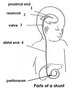
What are the parts of a shunt?
Most shunts have 4 parts:
1. The proximal end. It is put into the ventricle. There are many small holes on this end.
2. A reservoir. This lets the shunt pressure be checked. It is also used to get samples of CSF.
3. A valve. This allows for one-way flow of CSF. Some valves can be adjusted for different opening pressures.
4. The distal end. It helps get the CSF to a place where is can be absorbed. The end is put into the:
- Peritoneum (VP), such as the atrium of the heart (VA), or
- The pleural cavity of the lung (VPI).
Special instructions
- A shunt-o-gram should not be done if your child has a fever.
- If your child has a hard time lying still or is upset about the procedure, let the radiology staff know ahead of time.
- If your child cannot lie still for the scan, they may need special medicine called sedation. This helps them stay still. Do not let your child eat or drink anything before the scan. Use the guide below:
No solid food 8 hours before the scan.
No milk or formula for 6 hours before the scan.
No breast milk for 4 hours before the scan.
No clear liquids for 2 hours before the scan.
Please note: It is important that you follow these special instructions. If your child eats or drinks anything after the times listed above, the scan may be cancelled.
How the test is done
- When it is time for the scan, your child will go with the nuclear medicine tech to radiology. Your child will lie on their back on a special table in the exam room.
- When your child is comfortable, a small amount of hair will be shaved off over the shunt reservoir and the skin will be cleaned.
- A provider from the neurosurgery team will put a small needle in the shunt reservoir. Your child will feel a small prick in the skin. The pressure is measured and a small amount of CSF is removed. Some of the CSF may be sent to the lab to check for infection.
- A provider from the neurosurgery team will inject the isotope and the small amount of CSF that was removed back into the shunt reservoir. Pictures will be taken of the proximal end to see if there is flow into the ventricles of the brain.
- For a VP shunt, pictures of the distal end will be taken to see if the isotope flows to the abdomen.
- If flow is not seen after 10 minutes, the radiologist may pump the shunt and/or ask your child to sit up to see if there is a block in the tubing or the abdomen.
Results
The radiologist will let your child’s doctor know the results of the test. The neurosurgery team will give directions about any other care that is needed.


