In this section
Transposition of the great arteries (TGA)
What is TGA | Causes | Why a concern? | Symptoms | Diagnosis | Treatment | Postoperative care | Care at home | Long-term outlook | Find a doctor | Locations
 |
|
Ava was diagnosed with transposition of the great arteries/vessels. Read Ava's story. |
Dextro-Transposition of the great arteries (TGA or d-TGA) is a congenital heart defect (present at birth) in which the two main arteries of the heart are completely reversed from how the heart normally develops. Babies with a TGA heart defect are born with two separate paths of blood circulation, which doesn’t allow the blood to become enriched enough with oxygen before being pumped to the body. Other heart defects may be present, too, as the baby’s body adapts to survive with TGA.
Experts in treating transposition of the great arteries
Transposition of the great arteries can be life-threatening, so infants born with TGA will likely be admitted to the cardiac intensive care unit (CICU) immediately after delivery. Surgery is a required treatment for TGA and usually takes place within one or two weeks of birth. Ideally, babies will have been diagnosed with TGA before birth and delivered in a hospital with the expertise to care for babies with critical congenital heart defects.
The Herma Heart Institute’s pediatric cardiologists and heart surgeons have extensive experience in treating children with transposition of the great arteries and offer excellent surgical outcomes. Their expertise in all congenital heart defects that may impact your child, along with an onsite Birth Center and our advanced cardiac intensive care specialization, makes the Herma Heart Institute team a trusted partner in your child’s care.
The Herma Heart Institute consistently achieves outstanding outcomes for congenital heart surgery for even the most complex types of heart disease, as evaluated by the Society of Thoracic Surgeons. Learn more about our heart surgery outcomes.


Note: Data is provided from the Society of Thoracic Surgeons (STS). New data is not available beyond June 2019, as STS is in the process of refining their reporting methodologies. Hospitals are also limited in providing updated data due to the demands of the COVID-19 pandemic.
About transposition of the great arteries (TGA)

In babies with transposition of the great arteries, abnormal development of the fetal heart takes place during the first eight weeks of pregnancy. The large vessels that take blood away from the heart to the lungs, or to the body, are improperly connected.
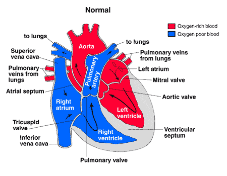
Normally, oxygen-poor (blue) blood returns to the right atrium from the body, travels to the right ventricle, then is pumped through the pulmonary artery into the lungs where it receives oxygen. Oxygen-rich (red) blood returns to the left atrium from the lungs, passes into the left ventricle, and then is pumped through the aorta out to the body.
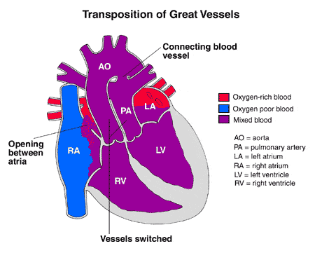
In transposition of the great arteries, the aorta is connected to the right ventricle, and the pulmonary artery is connected to the left ventricle - the exact opposite of a normal heart's anatomy.
- Oxygen-poor (blue) blood returns to the right atrium from the body, passes through the right atrium and ventricle, then goes into the misconnected aorta back to the body.
- Oxygen-rich (red) blood returns to the left atrium from the lungs, passes through the left atrium and ventricle, then goes into the pulmonary artery and back to the lungs.

Two separate circuits are formed - one that circulates oxygen-poor (blue) blood from the lungs back to the lungs, and another that recirculates oxygen-rich (red) blood from the body back to the body.
Other TGA heart defects
Other heart defects are often associated with TGA, and they actually may be necessary in order for an infant with transposition of the great arteries to live. An opening in the atrial or ventricular septum will allow blood from one side to mix with blood from another, creating "purple" blood with an oxygen level somewhere in-between that of the oxygen-poor (blue) and the oxygen-rich (red) blood. Patent ductus arteriosus (another type of congenital heart defect) will also allow mixing of oxygen-poor (blue) and oxygen-rich (red) blood through the connection between the aorta and pulmonary artery. The "purple" blood that results from this mixing is beneficial, providing at least smaller amounts of oxygen to the body, if not a normal amount of oxygen.
Because of the low amount of oxygen provided to the body, TGA is a heart problem that is labeled "blue-baby syndrome."
Transposition of the great arteries is the second most common congenital heart defect that causes problems in early infancy. TGA occurs in 5 to 7 percent of all congenital heart defects. Sixty to 70 percent of the infants born with the defect are boys.
What causes TGA Heart Defects?
The heart is forming during the first 8 weeks of fetal development. The problem occurs in the middle of these weeks, allowing the aorta and pulmonary artery to be attached to the incorrect chamber.
Some congenital heart defects may have a genetic link, either occurring due to a defect in a gene, a chromosome abnormality, or environmental exposure, causing heart problems to occur more often in certain families. Most of the time this heart defect occurs sporadically (by chance), with no clear reason for its development.
Why is TGA a concern?
Babies with TGA have two separate circuits - one that circulates oxygen-poor (blue) blood from the lungs back to the lungs, and another that recirculates oxygen-rich (red) blood from the body back to the body. Without an additional heart defect that allows mixing of oxygen-poor (blue) and oxygen-rich (red) blood, such as an atrial or ventricular septal defect, infants with TGA will have oxygen-poor (blue) blood circulating through the body - a situation that is fatal. Even with an additional defect present that allows mixing, babies with transposition of the great arteries will not have enough oxygen in the bloodstream to meet the body's demands.
Even when a good bit of mixing of oxygen-poor (blue) and oxygen-rich (red) blood can occur, other problems are present. The left ventricle, which in TGA is connected to the pulmonary artery, is the stronger of the two ventricles since it normally has to generate a lot of force to pump blood to the body. The right ventricle, connected to the aorta in TGA, is the weaker of the two ventricles. Because the right ventricle is weaker, it may not be able to pump blood efficiently to the body, and it will enlarge under the strain of the job. The left ventricle may pump blood into the lungs more vigorously than needed, leading to strain in the blood vessels in the lungs.
Signs and Symptoms of TGA
The obvious TGA symptom is a newborn who becomes cyanotic (blue) in the transitional first day of life when the maternal source of oxygen (from the placenta) is removed. Cyanosis is noted in the first hours of life in about half of the infants with TGA, and within the first days of life in 90 percent of them. The degree of cyanosis is related to the presence of other defects that allow blood to mix, including a patent ductus arteriosus - a fetal connection between the aorta and the pulmonary artery present in the newborn, which usually closes in the first few days after birth.
The following are the other most common TGA heart symptoms. However, each child may experience symptoms differently. Symptoms may include:
- Rapid breathing
- Labored breathing
- Rapid heart rate
- Cool, clammy skin
The symptoms of TGA may resemble other medical conditions or heart problems. Always consult your child's physician for a diagnosis.
How is TGA diagnosed?
A pediatric cardiologist and/or a neonatologist may be involved in your child's care. A pediatric cardiologist specializes in the diagnosis and medical management of congenital heart defects, as well as heart problems that may develop later in childhood. A neonatologist specializes in illnesses affecting newborns, both premature and full-term.
Cyanosis is the major indication that there is a problem with your newborn. Your child's physician may have also heard a heart murmur during a physical examination. A heart murmur is simply a noise caused by the turbulence of blood flowing through the openings that allow the blood to mix.
Other diagnostic tests are needed to help with the diagnosis, and may include the following:
- Chest x-ray - a diagnostic test which uses invisible electromagnetic energy beams to produce images of internal tissues, bones, and organs onto film.
- Electrocardiogram (ECG or EKG) - a test that records the electrical activity of the heart, shows abnormal rhythms (arrhythmias or dysrhythmias), and detects heart muscle stress.
- Echocardiogram (echo) - a procedure that evaluates the structure and function of the heart by using sound waves recorded on an electronic sensor that produce a moving picture of the heart and heart valves.
- Cardiac catheterization - a cardiac catheterization is an invasive procedure that gives very detailed information about the structures inside the heart. Under sedation, a small, thin, flexible tube (catheter) is inserted into a blood vessel in the groin, and guided to the inside of the heart. Blood pressure and oxygen measurements are taken in the four chambers of the heart, as well as the pulmonary artery and aorta. Contrast dye is also injected to more clearly visualize the structures inside the heart.
TGA Treatment
Specific treatment for transposition of the great arteries will be determined by your child's physician based on:
- Your child's age, overall health, and medical history
- Extent of the disease
- Your child's tolerance for specific medications, procedures, or therapies
- Expectations for the course of the disease
- Your opinion or preference
Your child will most likely be admitted to the intensive care unit (ICU) or special care nursery once symptoms are noted. Initially, your child may be placed on oxygen, and possibly even on a ventilator, to assist his/her breathing. Intravenous (IV) medications may be given to help the heart and lungs function more efficiently.
Other important aspects of initial treatment include the following:
- A cardiac catheterization procedure can be used as a diagnostic procedure, as well as initial treatment procedure for some heart defects. A cardiac catheterization procedure will usually be performed to evaluate the defect(s) and the amount of blood that is mixing.
- As part of the cardiac catheterization, a procedure called a balloon atrial septostomy may be performed to improve mixing of oxygen-rich (red) and oxygen-poor (blue) blood.
- A special catheter with a balloon in the tip is used to create an opening in the atrial septum (wall between the left and right atria).
- The catheter is guided through the foramen ovale (a small opening present in the atrial septum that closes shortly after birth) and into the left atrium.
- The balloon is inflated.
- The catheter is quickly pulled back through the hole, into the right atrium, enlarging the hole, allowing blood to mix between the atria.
- An intravenous medication called prostaglandin E1 is given to keep the ductus arteriosus from closing.
Within the first 1 to 2 weeks of age, transposition of the great arteries is surgically repaired. The procedure that accomplishes this is called a "switch," which roughly describes the surgical process.
The operation is performed under general anesthesia, and involves the following:
- The aorta is moved from the right ventricle to its normal position over the left ventricle.
- The pulmonary artery is moved from the left ventricle to its normal position over the right ventricle.
- The coronary arteries are moved so they will originate from the aorta and take oxygen-rich (red) blood to the heart muscle.
- Other defects, such as atrial or ventricular septal defects or a patent ductus arteriosus, are commonly closed.
TGA Heart - Postoperative care for your child
After surgery, infants will return to the intensive care unit (ICU) for a few days to be closely monitored during recovery.
While your child is in the ICU, special equipment will be used to help him/her recover, and may include the following:
- Ventilator - a machine that helps your child breathe while he/she is under anesthesia during the operation. A small, plastic tube is guided into the windpipe and attached to the ventilator, which breathes for your child while he/she is too sleepy to breathe effectively on his/her own. After a transposition of the great arteries, children will benefit from remaining on the ventilator overnight or even longer so they can rest.
- Intravenous (IV) catheters - small, plastic tubes inserted through the skin into blood vessels to provide IV fluids and important medicines that help your child recover from the operation.
- Arterial line - a specialized IV line is placed in the wrist or other area of the body where a pulse can be felt, that measures blood pressure continuously during surgery and while your child is in the ICU.
- Nasogastric (NG) tube - a small, flexible tube that keeps the stomach drained of acid and gas bubbles that may build up during surgery.
- Urinary catheter - a small, flexible tube that allows urine to drain out of the bladder and accurately measures how much urine the body makes, which helps determine how well the heart is functioning. After surgery, the heart will be a little weaker than it was before, and, therefore, the body may start to hold onto fluid, causing swelling and puffiness. Diuretics may be given to help the kidneys to remove excess fluid from the body.
- Chest tube - a drainage tube may be inserted to keep the chest free of blood that would otherwise accumulate after the incision is closed. Bleeding may occur for several hours, or even a few days after surgery.
- Heart monitor - a machine that constantly displays a picture of your child's heart rhythm, and monitors heart rate, arterial blood pressure, and other values.
Your child may need other equipment not mentioned here to provide support while in the ICU, or afterwards. The hospital staff will explain all of the necessary equipment to you.
Your child will be kept as comfortable as possible with several different medications; some which relieve pain, and some which relieve anxiety. The staff will also be asking for your input as to how best to soothe and comfort your child.
After being discharged from the ICU, your child will recuperate on another hospital unit for a few days before going home. You will learn how to care for your child at home before your child is discharged. Your child may need to take medications for a while, and these will be explained to you. The staff will give you written instructions regarding medications, activity limitations, and follow-up appointments before your child is discharged.
Infants who spent a lot of time on a ventilator, or who were fairly ill while in the ICU, may have trouble feeding initially. These babies may have an oral aversion; they might equate something placed in the mouth, such as a pacifier or bottle, with a less pleasant sensation such as being on the ventilator. Some infants are just tired, and need to build their strength up before they will be able to learn to bottle feed. Strategies used to help infants with nutrition include the following:
- High-calorie formula or breast milk - Special nutritional supplements may be added to formula or pumped breast milk that increase the number of calories in each ounce, thereby allowing your baby to drink less and still consume enough calories to grow properly.
- Supplemental tube feedings - Feedings given through a small, flexible tube that passes through the nose, down the esophagus, and into the stomach, that can either supplement or take the place of bottle-feedings. Infants who can drink part of their bottle, but not all, may be fed the remainder through the feeding tube. Infants who are too tired to bottle-feed at all may receive their formula or breast milk through the feeding tube alone.
Caring for your child at home following a TGA surgical repair
Pain medications, such as acetaminophen or ibuprofen, may be recommended to keep your child comfortable at home. Your child's physician will discuss pain control before your child is discharged from the hospital.
If any special treatments are to be given at home, the nursing staff will ensure that you are able to provide them, or a home health agency may assist you.
You may receive additional instructions from your child's physicians and the hospital staff.
Long-term outlook and prognosis after TGA heart surgical repair
Many infants who undergo TGA surgical repair will grow and develop normally. However, after TGA repair, your infant will need to be followed periodically by a pediatric cardiologist who will make assessments to check for any heart-related problems, which can include the following:
- Fast, slow, or irregular heart rhythms
- Leaky heart valves
- Narrowing of one or both of the great arteries at the switch connection site(s)
- Narrowing of the coronary arteries at their switch connection site
Consult your child's physician regarding the specific outlook for your child.
Let us help you
Coming from out of town?
Traveling with a sick child to a new city can be stressful. We can make your visit to our hospital as easy as possible.
Traveling here locally?
Contact us for more information about the Herma Heart Institute. Request an appointment online or call (414) 607-5280 or toll-free (877) 607-5280.
