Retinoblastoma Glossary
Glossary
Bilateral retinoblastoma: A cancerous tumor(s) located in the retina of both eyes.
Brachytherapy: A cancer treatment where radioactive seeds are placed near or inside a tumor.
Chemotherapy: A cancer treatment that uses drugs to destroy cancer cells or to stop their growth. It can be given many ways: orally, through an IV infusion or by injection. Chemotherapy works by interfering with rapid growth and division of cancer cells. It can also affect healthy rapidly dividing cells. These drugs disrupt the cell cycle and prevent cancer cells from multiplying This causes them to be destroyed.
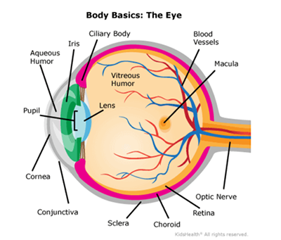
Choroid: The middle layer of the eye which contains blood vessels to nourish the eye.
Conformer: A temporary, oval shaped plastic device. It is used after an eye removal surgery (enucleation) to help maintain the shape of the eye socket. It also allows the eyelids to function normally while the eye socket is healing.
Cryotherapy: Treatment that uses extreme cold to freeze and destroy small retinoblastoma tumors.
Enucleation: Surgical removal of the eye.
Fundus Drawings: Map of the eye with tumor sketches drawn by an ophthalmologist.
Intra-arterial Chemotherapy (IAC): Method of delivering a concentrated dose of cancer-destroying medicine (chemotherapy) directly to the eye, with minimal systemic toxicity.
Intra-vitreal Injection (IVC): Injection of medicine through the wall of the eye into the vitreous.
Iris: The colored part of the eye that surrounds the pupil.
Laser therapy: Light therapy consisting of laser beams used to treat small retinoblastoma tumors(s).
Leukocoria: Causes the pupil of the eye to reflect white instead of the normal black (or red reflection in a flash photograph).
Macula: The area of the retina that is responsible for central vision. It houses the eyes rods (sensitive to light, responsible for might vision and peripheral vision) and cones (responsible for sharp vision and color).
MRI (Magnetic Resonance Imaging): A non-invasive test which uses a strong magnetic field and radio waves. It creates dilated images to view the eye and brain without radiation exposure.
Optic Nerve: Contains nerve fibers which send info to the brain for interpretation of objects seen. It contains about a million cells.
Prosthesis: An artificial eye designed to restore appearance and to support the eye socket after an eye is removed.
Pupil: Black, circular opening in the center of the eyes. It is the opening through which light enters the eye. This allows a person to see.
Radioactive seeds: Small radioactive pellets used for a type of cancer treatment call brachytherapy. The seeds are placed near a tumor to deliver a high dose of radiation directly to the cancer cells, minimizing exposure to surrounding healthy tissues.
Retina: The light sensitive tissue of the inner layer of the eye consisting of a thin membranous lining. The retina is what allows you to see. It has 10 layers and contains millions of cells. It has nerves that bring information to the brain from the optic nerve for seeing. This is where retinoblastoma starts.
Retinoblastoma: The most common type of eye cancer in children. There are approximately 300 newly diagnosed cases a year in the United States. Retinoblastoma happens most often in children under the age of 5 years old. It affects children of all races, boys, and girls equally. The tumor may be in 1 eye (unilateral) or in both eyes (bilateral). The average age of diagnosis is 2.5 years old if one eye is affected and 1 years old if both eyes are affected.
Sclera: Outer protective white coating of the eye.
Strabismus: The second most common sign of retinoblastoma. The child’s eye turns inward towards the nose or outward towards the ear.
Systemic Chemotherapy: A cancer treatment where anti-cancer drugs (chemotherapy) travel throughout the blood stream to reach and destroy cancer cells throughout the body. This treatment is given by IV infusion, injection or orally.
Unilateral Retinoblastoma: A cancerous tumor(s) located in the retina of 1 eye.
Vitreous: A clear, gel-like substance. It makes up the largest part of the eye. It fills the space between the lens and the retina. Its function is to maintain the eyes spherical shape, allowing light to pass through to the retina and helps to absorb shock.
© 2025 The Nemours Foundation/KidsHealth®. Used and adapted under license by Children’s Wisconsin. This information is for general use only. For specific medical advice or questions, consult your health care professional.
Draft Number: 2242