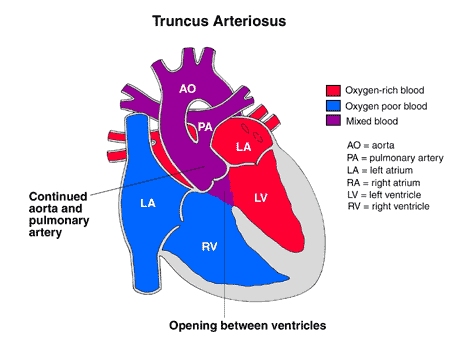
Truncus arteriosus

About truncus arteriosus | Causes | Why a concern? | Symptoms | Diagnosis | Treatment | Postoperative care | Care at home | Long-term outlook | Find a doctor | Locations

 Little boy that had Truncus Arteriosus surgery riding a bike.
Gavin was born with truncus arteriosus, a rare heart defect. Read Gavin's story.

Truncus arteriosus is a rare and complex congenital heart defect (present at birth) that develops during the first eight weeks of pregnancy. Truncus arteriosus occurs when the two main blood vessels, the aorta and the pulmonary artery, stay connected and fail to separate completely as the fetus develops. Truncus arteriosus is further complicated because another congenital heart defect, ventricular special defect (VSD), occurs with it.

In babies born with truncus arteriosus the blood passing through their systems is poor in oxygen, causing difficulty breathing, rapid heart rate, varying degrees of cyanosis (blue color of the skin, lips and nailbeds), and other symptoms. Corrective surgery is required, usually within one or two weeks. Most babies born with truncus arteriosus do well, but will need lifelong monitoring so that they can continue to live long, healthy lives.

Experts in truncus arteriosus treatment

Get a second opinion

Truncus arteriosus is a complex, serious condition that requires surgery and may result in additional procedures as the child matures. The Herma Heart Institute’s pediatric cardiologists and heart surgeons are highly experienced in treating children with truncus arteriosus and other complex congenital defects, and offer excellent surgical outcomes for truncus arteriosus repair. Our on-site Birth Center, advanced cardiac intensive care expertise and family-focused approach makes the Herma Heart Institute team a trusted partner in your child’s care.

The Herma Heart Institute consistently achieves outstanding outcomes for congenital heart disease for even the most complex types of heart disease. Learn more about our heart surgery outcomes.
Truncus arteriosus

Note: Data is provided from the Society of Thoracic Surgeons (STS). New data is not available beyond June 2019, as STS is in the process of refining their reporting methodologies. Hospitals are also limited in providing updated data due to the demands of the COVID-19 pandemic.

A diagram of a normal heart.

What is truncus arteriosus?

Typically, the heart begins as a hollow tube, and the chambers, valves, and great arteries develop throughout the first eight weeks of pregnancy. The aorta and pulmonary artery start as a single blood vessel, which eventually divides and becomes two separate arteries. Truncus arteriosus occurs when the single great vessel fails to separate completely, leaving a connection between the aorta and pulmonary artery.

Truncus arteriosis is a complex defect where there is a single (normally there are two separate arteries) vessel arising from the heart that forms the aorta and pulmonary artery. Another congenital heart defect that occurs with truncus arteriosus is a ventricular septal defect (a flaw in the ventricular septum, or dividing wall between the two lower chambers of the heart known as the right and left ventricles).

Normally, there are two separate arteries (the aorta and the pulmonary artery). Oxygen-poor (blue) blood returns to the right atrium from the body, travels to the right ventricle, then is pumped through the pulmonary artery into the lungs where it receives oxygen. Oxygen-rich (red) blood returns to the left atrium from the lungs, passes into the left ventricle, and then is pumped through the aorta out to the body.

Diagram of a heart with truncus arteriosus heart condition. 

In truncus arteriosus, oxygen-poor (blue) and oxygen-rich (red) blood mix back and forth through the ventricular septal defect. This mixed blood then flows through the common truncal vessel. Some of it will flow through the branch that becomes the pulmonary artery and on to the lungs, and some of the mixed blood will go into the aortic branch and continue to the body. The mixed blood that goes to the body does not have as much oxygen as normal, and will cause varying degrees of cyanosis (blue color of the skin, lips, and nailbeds).

Truncus arteriosus occurs in less than one out of every 10,000 live births. It makes up 1 percent of all cases of congenital heart disease.

Causes of truncus arteriosus

Some congenital heart defects may have a genetic link, either occurring due to a defect in a gene, a chromosome abnormality, or environmental exposure, causing heart problems to occur more often in certain families. Other times this heart defect occurs sporadically (by chance), with no clear reason for its development.

Why is truncus arteriosus a concern?

The blood that passes through the common truncal vessel has a lower oxygen content than normal. Oxygen-poor (blue) blood from the right ventricle and oxygen-rich (red) blood from the left ventricle mix together before entering the common vessel. Some of this mixed blood will go into the aorta and on to the body, producing cyanosis (blue color of the skin, lips, and nailbeds).

The pulmonary artery section of the common vessel gets more blood flow than the aorta does, because the pressure is lower in the lungs than the body and it is easier for blood to travel in that direction. If not repaired, the blood vessels in the lungs become damaged by the extra blood flow. As the pressure in the blood vessels in the lungs becomes higher, less blood goes to the lungs and more goes to the body. Cyanosis becomes worse as blood with lower amounts of oxygen travels to the body.

What are the symptoms of truncus arteriosus?

The following are the most common symptoms of truncus arteriosus. However, each child may experience symptoms differently. Symptoms may include:

  • Cyanosis
  • Fatigue
  • Sweating
  • Pale skin
  • Cool skin
  • Rapid breathing
  • Heavy breathing
  • Rapid heart rate
  • Congested breathing
  • Disinterest in feeding, or tiring while feeding
  • Poor weight gain

The symptoms of truncus arteriosus may resemble other medical conditions or heart problems. Always consult your child's physician for a diagnosis.

How is truncus arteriosus diagnosed?

Your child's physician may have heard a heart murmur during a physical examination, and referred your child to a pediatric cardiologist for a diagnosis. A heart murmur is simply a noise caused by the turbulence of blood flowing through the heart defects. Symptoms your child exhibits will also help with the diagnosis.

A pediatric cardiologist specializes in the diagnosis and medical management of congenital heart defects, as well as heart problems that may develop later in childhood. The cardiologist will perform a physical examination, listening to the heart and lungs, and make other observations that help in the diagnosis. The location within the chest that the murmur is heard best, as well as the loudness and quality of the murmur (harsh, blowing, etc.) will give the cardiologist an initial idea of which heart problem your child may have. However, other tests are needed to help with the diagnosis, and may include the following:

  • Chest x-ray - a diagnostic test which uses invisible electromagnetic energy beams to produce images of internal tissues, bones, and organs onto film.
  • Electrocardiogram (ECG or EKG) - a test that records the electrical activity of the heart, shows abnormal rhythms (arrhythmias or dysrhythmias), and detects heart muscle stress.
  • Echocardiogram (echo) - a procedure that evaluates the structure and function of the heart by using sound waves recorded on an electronic sensor that produce a moving picture of the heart and heart valves.
  • Cardiac catheterization - a cardiac catheterization is an invasive procedure that gives very detailed information about the structures inside the heart. Under sedation, a small, thin, flexible tube (catheter) is inserted into a blood vessel in the groin, and guided to the inside of the heart. Blood pressure and oxygen measurements are taken in the four chambers of the heart, as well as the pulmonary artery and aorta. Contrast dye is also injected to more clearly visualize the structures inside the heart.

Truncus arteriosus treatment

Specific treatment for truncus arteriosus will be determined by your child's physician based on:

  • Your child's age, overall health and medical history
  • Extent of the condition
  • Your child's tolerance for specific medications, procedures or therapies
  • Expectations for the course of the condition
  • Your opinion or preference

Truncus arteriosus must be treated by surgical repair of the defects. However, medical support may be necessary until the best time for the operation to take place. Treatment may include:

  • Medical management - Many children will eventually need to take medications to help the heart and lungs work better. Medication that may be prescribed includes the following:
    • Digoxin - a medication that helps strengthen the heart muscle, enabling it to pump more efficiently.
    • Diuretics - the body's water balance can be affected when the heart is not working as well as it could. These medications help the kidneys remove excess fluid from the body.
    • ACE (angiotensin-converting enzyme) inhibitors - dilates the blood vessels, making it easier for the heart to pump blood forward into the body. 
  • Adequate nutrition - Infants may become tired when feeding, and may not be able to eat enough calories to gain weight. Options that can be used to ensure your baby will have adequate nutrition include: 
    • High-calorie formula or breast milk - Special nutritional supplements may be added to formula or pumped breast milk that increase the number of calories in each ounce, thereby allowing your baby to drink less and still consume enough calories to grow.
    • Supplemental tube feedings - Feedings given through a small, flexible tube that passes through the nose, down the esophagus, and into the stomach, can either supplement or take the place of bottle feedings. Infants who can drink part of their bottle, but not all, may be fed the remainder through the feeding tube. Infants who are too tired to bottle feed may receive their formula or breast milk through the feeding tube alone. 
  • Surgical repair - Surgery is usually performed after the infant is 2 weeks old, but before the blood vessels in the lungs are overwhelmed by extra blood flow and become diseased. The operation is performed under general anesthesia, and involves the following:
    • The pulmonary arteries are detached from the common artery (truncus arteriosus) and connected to the right ventricle using a homograft (a section of pulmonary artery with its valves intact from a tissue donor).
    • The ventricular septal defect is closed with a patch. 

Postoperative care for your child

Children will spend time in the intensive care unit (ICU) after a truncus repair.

While your child is in the ICU, special equipment will be used to help him/her recover, and may include the following:

  • Ventilator - a machine that helps your child breathe while he/she is under anesthesia during the operation. A small, plastic tube is guided into the windpipe and attached to the ventilator, which breathes for your child while he/she is too sleepy to breathe effectively on his/her own. After a truncus repair, children will benefit from remaining on the ventilator overnight or even longer so they can rest.
  • Intravenous (IV) catheters - small, plastic tubes inserted through the skin into blood vessels to provide IV fluids and important medicines that help your child recover from the operation.
  • Arterial line - a specialized IV placed in the wrist or other area of the body where a pulse can be felt, that measures blood pressure continuously during surgery and while your child is in the ICU.
  • Nasogastric (NG) tube - a small, flexible tube that keeps the stomach drained of acid and gas bubbles that may build up during surgery.
  • Urinary catheter - a small, flexible tube that allows urine to drain out of the bladder and accurately measures how much urine the body makes, which helps determine how well the heart is functioning. After surgery, the heart will be a little weaker than it was before, and, therefore, the body may start to hold onto fluid, causing swelling and puffiness. Diuretics may be given to help the kidneys to remove excess fluid from the body.
  • Chest tube - a drainage tube may be inserted to keep the chest free of blood that would otherwise accumulate after the incision is closed. Bleeding may occur for several hours, or even a few days after surgery.
  • Heart monitor - a machine that constantly displays a picture of your child's heart rhythm, and monitors heart rate, arterial blood pressure, and other values. 

Your child may need other equipment not mentioned here to provide support while in the ICU, or afterwards. The hospital staff will explain all of the necessary equipment to you.

Your child will be kept as comfortable as possible with several different medications; some which relieve pain, and some which relieve anxiety. The staff will also be asking for your input as to how best to soothe and comfort your child.

After being discharged from the ICU, your child will recuperate in another hospital unit for a few days before going home. You will learn how to care for your child at home before your child is discharged. Your child may need to take medications for a while at home, and these will be explained to you. The staff will give you instructions regarding medications, activity limitations, and follow-up appointments before your child is discharged.

Caring for your child at home following truncus arteriosus repair

Pain medications, such as acetaminophen or ibuprofen, may be recommended to keep your child comfortable at home. Your child's physician will discuss pain control before your child is discharged from the hospital.

Often, infants who fed poorly prior to surgery have more energy after the recuperation period, and begin to eat better and gain weight faster. However, high-calorie formulas may be needed for several weeks or months after surgery to help your child catch up growth-wise. Tube feedings may also be helpful until your child is able to feed better.

After surgery, older children usually have a fair tolerance for activity. Your child may become tired easily, and sleep more right after surgery, but, within a few weeks, your child should be fully recovered.

You may receive additional instructions from your child's physicians and the hospital staff.

Long-term outlook after truncus arteriosus repair

Many children who have had truncus arteriosus surgical repair can live healthy lives. Activity levels, appetite, and growth will eventually return to normal in most children.

Future intervention may be necessary if the pulmonary artery branches were small and do not grow well after surgery. The homograft connecting the right ventricle to the pulmonary artery may also need to be replaced in the future as your child grows.

Your child's cardiologist may recommend that antibiotics be given to prevent bacterial endocarditis after discharge from the hospital.

Consult your child's physician regarding the specific outlook for your child.

Choosing a heart hospital?

A young boy being tested for potential pediatrics heart conditions. He is holding his arms up and smiling.

Find tips for deciding whom to trust with your child’s heart surgery in a blog written by Peter Frommelt, MD, pediatric cardiologist and professor at the Medical College of Wisconsin.

Let us help you

Coming from out of town?
Traveling with a sick child to a new city can be stressful. We can make your visit to our hospital as easy as possible.
Traveling here locally?
Contact us for more information about the Herma Heart Institute. Request an appointment online or call (414) 607-5280 or toll-free (877) 607-5280.