
In this section
Hydrocephalus
What is hydrocephalus?
The brain and spinal cord are surrounded by cerebral spinal fluid. Small chambers in the brain are called ventricles. Normally, cerebral spinal fluid is produced primarily in the two lateral ventricles. Then, the cerebral spinal fluid drains through the third and fourth ventricles and is circulated around the brain and spinal cord.
This cerebral spinal fluid acts as a cushion for the brain and is reabsorbed by the membrane covering the brain. With hydrocephalus, the cerebral spinal fluid backs up in the ventricles, causing them to expand and push on the surrounding brain.
This increased size in the ventricles may be referred to as ventriculomegaly. Ventriculomegaly occurs when the two lateral ventricles are greater than 1 cm in width but less than 1.5 cm. Sometimes ventriculomegaly will regress or return to normal size and there is no long-term problem. Hydrocephalus occurs when the two lateral ventricles are greater than 1.5 cm in width. The ventricles can fill to such an extent that the fetus’s head size becomes enlarged.
What causes hydrocephalus?
The major causes of hydrocephalus include:
- The flow of cerebral spinal fluid is blocked
- Abnormal circulation of cerebral spinal fluid causes inadequate absorption by the membranes covering the brain
- The brain size is small (cerebral atrophy or there is a localized injury), and the fluid volume appears large as it fills in space
- Very rarely, it may be inherited
Cerebral spinal fluid production normally increases late in gestation, so the ventricles and head size need close monitoring to determine whether there is really a concern. Ventriculomegaly may be the first sign of another anomaly, either with the central nervous system or outside the central nervous system.
Experts estimate that 50 to 80 percent of babies with hydrocephalus will have another anomaly outside the central nervous system, and approximately a third will have an associated anomaly within the central nervous system.
Hydrocephalus occurs more frequently in males (64 percent males versus 36 percent females). It occurs in approximately 1 out of every 2,000 live births.
Prenatal diagnosis of hydrocephalus
If your doctor suspects that your baby has hydrocephalus, he or she may order additional tests, including:
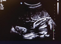
- Ultrasound: Hydrocephalus may be diagnosed on a routine ultrasound. The ventricles within the head will measure larger than normal. The head circumference may remain within the normal limits, or it may become enlarged as the pregnancy progresses. Your obstetrician will likely refer you to a perinatologist (a specialist who handles high-risk pregnancies). Your perinatologist will perform a targeted ultrasound to examine the anatomy of the brain to confirm ventriculomegaly. He or she will also look for any associated anomalies.
- Amniocentesis: Hydrocephalus is occasionally associated with chromosomal abnormalities. Your perinatologist will typically offer an amniocentesis to look for chromosomal issues.
- MRI: Your doctor may also recommend another a fetal MRI. Ultrasound imaging can be limited by your body, the surrounding amniotic fluid and the position of your baby in the womb. Fetal MRI is a non-invasive diagnostic test that produces better images of soft tissue, and bone or dense tissue does not interfere with the image. The biggest limitation of fetal MRI is that the best images are obtained when the fetus stays still, which can be difficult.
How does hydrocephalus affect my baby?
The amount of pressure on the brain from the increased size of the ventricles can alter the effect hydrocephalus has on your baby. Most children with simple hydrocephalus (no associated anomalies) that is diagnosed and treated early will function normally. Approximately 75 percent of children with hydrocephalus will have some form of motor disability. Many will have some type of learning disability.
How does hydrocephalus affect my pregnancy?
With the finding of ventriculomegaly on a routine ultrasound, your physician may refer you to a perinatologist. The perinatologist will perform a targeted ultrasound to examine the anatomy of the brain to confirm ventriculomegaly.
Your perinatologist will also look for any associated anomalies, which have been reported in 54 to 84 percent of hydrocephalus cases. These associated anomalies may be disorders within the brain (intracranial) or outside of the brain (extracranial). Between 12 and 25 percent of cases also have an associated genetic disorder.
It is important to rule out an intrauterine infection such as cytomegalovirus, toxoplasmosis, syphilis, intracranial hemorrhage and tumor of the central nervous system. These causes of hydrocephalus typically lead to increased size in the lateral ventricles, but not an increase in the head size. This is because the cerebral spinal fluid is filling in space from damage caused to the brain from the infection or injury. Often, these babies will have microcephaly or a smaller than average size head.
Extracranial abnormalities have been seen in as many as 2/3rds of babies with hydrocephalus. The major extracranial defect associated with hydrocephalus is mylomeningocele.
Type of delivery
Your baby's head growth and ventricle size will be monitored closely throughout the remainder of your pregnancy. The method of delivery will be determined by your baby's head size and his or her well-being at the time of labor and delivery, as with any pregnancy. Hydrocephalus does not, by itself, necessitate a Caesarian section delivery.
How do you treat hydrocephalus?
Treatment will be necessary if the ventricles continue to expand and the head circumference grows too fast. Your baby will be evaluated after birth for the need of a shunt. If a shunt is deemed necessary, your baby will need surgery. A shunt is a thin tube. One end of this tube is placed in the ventricle, and the other end is passed underneath the skin and drains into the abdominal cavity where the body can reabsorb the cerebral spinal fluid. As your baby grows, the shunt will need to be replaced approximately every 2 to 4 years.
If we believe that your baby’s ventricles are large because of severe brain damage, we can openly discuss other treatment options, such as palliative care.
What happens after surgery?
If a shunt is necessary, your baby will need surgery. After surgery, your baby may need some help breathing. In that case, we will place a special tube that may stay in place until your baby is able to breathe effectively on his or her own.
Placing IV and other lines: We may also place some special IV lines. The umbilical cord normally has two arteries and one vein. We may place a line in the vein as a means to provide nourishment until your baby is able to eat. And we may place another line in one of the arteries. This is called an arterial line. Through this line we can:
- Give fluids and medication
- Monitor blood pressure monitored
- Draw blood for lab work
Providing nutrition: Until your baby is able to eat, he or she will receive total parenteral nutrition (TPN). This is an IV solution that contains protein, fats, sugar, vitamins, and mineral. It will supply all your baby's nutritional requirements until he or she is able to take food by mouth.
Observing and monitoring your baby: We will observe your baby for infection and proper shunt drainage. We will also monitor his or her head circumference.
You will learn how to care for, feed and monitor your baby in preparation for taking him or her home. For example, sometimes the shunt can become clogged or may stop draining effectively, which causes an increase in the pressure inside your baby's head. You will be trained to recognize signs and symptoms of this and other potential complications.
Will I be able to care for my baby?
Yes. Please ask your baby's nurse about ways to interact with and care for your baby.
If you had planned to breastfeed your baby, you can begin to pump and freeze breast milk while you are still in the hospital. A lactation consultant can answer your questions. Your milk will be frozen and stored in the Neonatal Intensive Care Unit (NICU) until your baby is ready for it. The NICU has breast pumps and private rooms available to you when you are visiting.
You can bring in pictures, small toys, booties and blankets for your baby while he or she is in the NICU.
When can my baby go home?
Your baby will go home when he or she is eating and tolerating enough food to grow and gain weight. A baby with simple hydrocephalus that requires shunt placement may be home within 2 weeks.
If your baby has any other complicating defects, his or her stay will be prolonged according to the severity of those anomalies.
If you have decided to pursue palliative care for your baby, you also have the option of taking your infant home with the support of hospice services. If necessary, we will discuss this option with you.
What is my baby's long-term prognosis?
Prognosis is dependent on any associated defects that may be related to the cause of hydrocephalus, as well as the amount of pressure exerted on the brain tissue prior to shunting.
The effects of hydrocephalus vary and partly depend upon the cause and treatment required, as well as your baby's response to treatment.
The key to a good prognosis is early detection and treatment, along with preventing infections. Most of the newborns born with hydrocephalus will have a normal lifespan, and approximately 40 to 50 percent will have normal intelligence. Seizure disorders have been diagnosed in about 10 percent of children with hydrocephalus. The mortality rate for infants is approximately 5 percent.
Studies have shown that the risk of shunt failure in the infant's first year is 30 percent. Shunts, on average, are revised about two times in the first 10 years. The risk of intellectual disability is approximately 35 percent. Your baby's pediatrician and pediatric neurologist will provide follow-up care once your baby is home.
Learn more about the craniofacial program at Children's Wisconsin.
Get a second opinion
(414) 240-1831
Research and outcomes
Our outcomes reports help families and partner providers make the most informed healthcare decisions. Learn more about our surgical outcomes and current research studies.