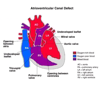
Atrioventricular canal defect (AV canal defect)

About atrioventricular canal | Causes | Why a concern? | Symptoms | Diagnosis | Treatment | Postoperative care | Care at home | Long-term outlook | Find a doctor | Locations

What is an atrioventricular canal defect?

Atrioventricular canal (AVC, or AV canal) is a combination of congenital (present at birth) heart defects that together create a large defect inside the heart. As the fetus is growing, something occurs to affect the heart’s development during the first eight weeks of pregnancy and some areas of the heart do not form properly. The result is several related abnormalities of the heart’s structures that most often require surgical treatment.

Experts in AV canal defect treatment

Get a second opinion

The development of AV canal defect (AVC) is rare and often linked to chromosome abnormalities such as those associated with Down’s syndrome. The surgery required to correct the condition usually occurs before a child is 6 months old. Left untreated, AV canal defect can lead to lung disease and serious infections in the heart (bacterial endocarditis). Herma Heart Institute’s pediatric cardiologists and heart surgeons have extensive experience in treating children with atrioventricular canal defects and offer excellent surgical outcomes. Additionally, they partner in care with other experts your child may need, such as those leading the Genetics Program and the Down Syndrome Clinic at Children's Wisconsin.

The Herma Heart Institute consistently outperforms when it comes to congenital heart surgery outcomes for even the most complex types of heart disease, as evaluated by the Society of Thoracic Surgeons. Learn more about our heart surgery outcomes.
AV canal repair

Note: Data is provided from the Society of Thoracic Surgeons (STS). New data is not available beyond June 2019, as STS is in the process of refining their reporting methodologies. Hospitals are also limited in providing updated data due to the demands of the COVID-19 pandemic.

About AV canal defects

Atrioventricular canal defect is a heart problem that involves several abnormalities of structures inside the heart, including the following:

  • Atrial septal defect - an opening in the atrial septum, or dividing wall between the two upper chambers of the heart known as the right and left atria.
  • Ventricular septal defect - an opening in the ventricular septum, or dividing wall between the two lower chambers of the heart known as the right and left ventricles.
  • Improperly formed mitral and/or tricuspid valves - the valves that separate the upper heart chambers (atria) from the lower heart chambers (ventricles) are improperly formed. 

Normally, oxygen-poor (blue) blood returns to the right atrium from the body, travels to the right ventricle, then is pumped into the lungs where it receives oxygen. Oxygen-rich (red) blood returns to the left atrium from the lungs, passes into the left ventricle, and then is pumped out to the body through the aorta.

An atrial septal defect allows oxygen-rich (red) blood to pass from the left atrium, through the opening in the septum, and then mix with oxygen-poor (blue) blood in the right atrium.

A ventricular septal defect allows oxygen-rich (red) blood to pass from the left ventricle, through the opening in the septum, and then mix with oxygen-poor (blue) blood in the right ventricle.

Illustration showing Atrioventricular Canal Defect. 

Abnormalities of the mitral or tricuspid valves allow blood that should be moving forward from the ventricle into either the pulmonary artery or the aorta to instead flow backward into the atria.

AV canal defect occurs in two out of every 10,000 live births, and equally in boys and girls.

What causes AV canal defect?

The heart is forming during the first 8 weeks of fetal development. It begins as a hollow tube, then partitions within the tube develop that eventually become the septa (or walls) dividing the right side of the heart from the left. Atrial and ventricular septal defects occur when the partitioning process does not occur completely, leaving openings in the atrial and ventricular septum. The valves that separate the upper and lower heart chambers are being formed in the latter portion of this eight-week period, and, for some reason, they do not develop properly, either.

There may be genetic influences on the development of atrioventricular canal. Consider the following statistics:

  • CHD is present in half of children born with Down syndrome, and 45 percent of these cases have AV canal defect. Down syndrome is caused by the presence of three #21 chromosomes in the cells of the body, rather than the usual pair (two) of chromosomes.
  • Similarly, one-third of all children born with atrioventricular canal defect also have Down syndrome.
  • Fourteen percent of mothers with an AV canal defect give birth to a child with the disease.
  • Some studies have investigated a possible link between the use of retinoic acid (a medication used for the treatment of dermatologic problems such as acne and facial wrinkles) while pregnant and the development of atrioventricular canal in the fetus. 

Other chromosome abnormalities (in addition to Down syndrome) are linked to the development of AV canal defect. Maternal age can have an effect on the prevalence of AV canal, which may be related to the increased probability of a woman giving birth to a child with Down syndrome as she gets older.

Why is AV canal defect a concern?

If not treated, this heart defect can cause lung disease. When blood passes through both the tand VSD from the left side of the heart to the right side, then a larger volume of blood than normal must be handled by the right side of the heart. This extra blood then passes through the pulmonary artery into the lungs, causing higher pressure than normal in the blood vessels in the lungs.

The larger the volume of blood that goes to the lungs, the higher the pressure. The lungs are able to cope with this extra pressure for a while, depending on exactly how high the pressure is. After a while, however, the blood vessels in the lungs become damaged by the extra pressure.

As pressure builds up in the lungs, the flow of blood from the left side of the heart to the right side and on to the lungs will diminish. This helps preserve the function of the lungs, but causes yet another problem. Blood flow within the heart goes from areas where the pressure is high to areas where the pressure is low. If the septal defects are not repaired, and lung disease begins to occur, pressure in the right side of the heart will eventually exceed pressure in the left. In this instance, it will be easier for oxygen-poor (blue) blood to flow from the right side of the heart, through the ASD and VSD, into the left ventricle, and on to the body. When this happens, the body does not receive enough oxygen in the bloodstream to meet its needs, and children may become cyanotic, or exhibit a blue coloring in their skin, lips, and nailbeds.

Because blood is pumped at high pressure through the septal openings, the lining of the right and left ventricles will become irritated and inflamed. Bacteria in the bloodstream can easily infect this injured area, causing a serious illness known as bacterial endocarditis.

What are atrioventricular canal defect symptoms?

The size of the septal openings will affect the type of atrioventricular canal defect symptoms noted, the severity of symptoms, and the age at which they first occur. The larger the openings, the greater the amount of blood that passes through from the left side of the heart to the right and overloads the right heart and the lungs.

Symptoms occur in infancy. The following are the most common symptoms of AV canal defect. However, each child may experience symptoms differently. Atrioventricular canal defect symptoms may include:

  • Fatigue
  • Sweating
  • Pale skin
  • Cool skin
  • Rapid breathing
  • Heavy breathing
  • Rapid heart rate
  • Congested breathing
  • Disinterest in feeding, or tiring while feeding
  • Poor weight gain

As the pressure in the lungs rises, blood within the heart will eventually "shunt" through the septal openings from the right heart to the left. This allows oxygen-poor (blue) blood to reach the body, and cyanosis will be noted. Cyanosis gives a blue color to the lips, nailbeds, and skin. The symptoms of AV canal defect may resemble other medical conditions or heart problems. Always consult your child's physician for a diagnosis.

How is AV canal defect diagnosed?

Your child's pediatrician may have heard a heart murmur during a physical examination, and referred your child to a pediatric cardiologist for a diagnosis. A heart murmur is simply a noise caused by the turbulence of blood flowing through the opening from the left side of the heart to the right. Symptoms your child exhibits will also help with the diagnosis.

A pediatric cardiologist specializes in the diagnosis and medical management of congenital heart defects, as well as heart problems that may develop later in childhood. The cardiologist will perform a physical examination, listening to the heart and lungs, and make other observations that help in the diagnosis. The location within the chest that the murmur is heard best, as well as the loudness and quality of the murmur (harsh, blowing, etc.) will give the cardiologist an initial idea of which heart problem your child may have. However, other tests are needed to help with the diagnosis, and may include the following:

  • Chest x-ray - a diagnostic test which uses invisible electromagnetic energy beams to produce images of internal tissues, bones, and organs onto film.
  • Electrocardiogram (ECG or EKG) - a test that records the electrical activity of the heart, shows abnormal rhythms (arrhythmias or dysrhythmias), and detects heart muscle stress.
  • Echocardiogram (echo) - a procedure that evaluates the structure and function of the heart by using sound waves recorded on an electronic sensor that produce a moving picture of the heart and heart valves. An echo can show the pattern of blood flow through the septal openings, and determine how large the openings are, as well as how much blood is passing through them.
  • Cardiac catheterization - a cardiac catheterization is an invasive procedure that gives very detailed information about the structures inside the heart. Under sedation, a small, thin, flexible tube (catheter) is inserted into a blood vessel in the groin, and guided to the inside of the heart. Blood pressure and oxygen measurements are taken in the four chambers of the heart, as well as the pulmonary artery and aorta. Contrast dye is also injected to more clearly visualize the structures inside the heart. 

Atrioventricular canal defect treatment

Specific atrioventricular canal treatment will be determined by your child's physician based on:

  • Your child's age, overall health, and medical history
  • Extent of the disease
  • Your child's tolerance for specific medications, procedures, or therapies
  • Expectations for the course of the disease
  • Your opinion or preference.

AV canal defect is treated by surgical repair. However, medical support (i.e., medications) may be necessary until the operation is performed. Treatment may include:

  • Medical management - Many children will eventually need to take medications to help the heart and lungs work better, due to strain from the extra blood passing through the septal defects. Medications that may be prescribed include the following:
    • Digoxin - helps strengthen the heart muscle, enabling it to pump more efficiently.
    • Diuretics - the body's water balance can be affected when the heart is not working as well as it could. These medications help the kidneys remove excess fluid from the body.
    • ACE (angiotensin-converting enzyme) inhibitors - dilate the blood vessels, making it easier for the heart to pump blood forward into the body. 
  • Adequate nutrition - Infants may become tired when feeding, and may not be able to eat enough to gain weight. Options that can be used to ensure your baby will have adequate nutrition include: 
    • High-calorie formula or breast milk - Special nutritional supplements may be added to formula or pumped breast milk that increase the number of calories in each ounce, thereby allowing your baby to drink less and still consume enough calories to grow properly.
    • Supplemental tube feedings - Feedings given through a small, flexible tube that passes through the nose, down the esophagus, and into the stomach, can either supplement or take the place of bottle feedings. Infants who can drink part of their bottle, but not all, may be fed the remainder through the feeding tube. Infants who are too tired to bottle feed may receive their formula or breast milk through the feeding tube alone. 
  • Infection control - Children with certain heart defects are at risk for developing an infection of the inner surfaces of the heart known as bacterial endocarditis. A common procedure that puts your child at risk for this infection is a routine dental check-up and teeth cleaning. Other procedures may also increase the risk of the heart infection occurring. However, giving children with heart defects an antibiotic by mouth before these procedures can help prevent bacterial endocarditis. It is important that you inform all medical personnel that your child has a an atrioventricular canal defect so they may determine if the antibiotics are necessary before a procedure.
  • Surgical repair - The goal is to repair the septal openings and repair the valves before the lungs become damaged from too much blood flow and pressure. Your child's cardiologist will recommend when the repair should be performed based on results from the echocardiogram and cardiac catheterization. 
    • The operative methods used to repair AV canal defects have improved greatly in the past decade, and the operation has a high likelihood of success. Most children undergo surgery by the age of 6 months. Children with Down syndrome may develop lung problems earlier than other children, and may need to have surgical repair at an earlier age.
    • The operation is performed under general anesthesia.
    • The atrial and ventricular septal defects are often closed with a pericardial patch made from the membrane that covers the outside surface of the heart, or a patch made from synthetic material. The valve repair technique will differ according to the abnormality and the surgeon's preferences. 

AV Canal Surgery - Postoperative care for your child

Children will spend time in the cardiac intensive care unit (CICU) after an AV canal repair. During the first several hours after surgery, your child will be very drowsy from the anesthesia that was used during the operation, and from medications given to relax him/her and to help with pain. To help your child rest, these medications might be continued overnight or longer. As time goes by, your child will become more alert.

While your child is in the CICU, special equipment will be used to help him/her recover, and may include the following:

  • Ventilator - a machine that helps your child breathe while he/she is under anesthesia during the operation. A small, plastic tube is guided into the windpipe and attached to the ventilator, which breathes for your child while he/she is too sleepy to breathe effectively on his/her own. After a truncus repair, children will benefit from remaining on the ventilator overnight or even longer so they can rest.
  • Intravenous (IV) catheters - small, plastic tubes inserted through the skin into blood vessels to provide IV fluids and important medicines that help your child recover from the operation.
  • Arterial line - a specialized IV placed in the wrist or other area of the body where a pulse can be felt, that measures blood pressure continuously during surgery and while your child is in the ICU.
  • Nasogastric (NG) tube - a small, flexible tube that keeps the stomach drained of acid and gas bubbles that may build up during surgery.
  • Urinary catheter - a small, flexible tube that allows urine to drain out of the bladder and accurately measures how much urine the body makes, which helps determine how well the heart is functioning. After surgery, the heart will be a little weaker than it was before, and, therefore, the body may start to hold onto fluid, causing swelling and puffiness. Diuretics may be given to help the kidneys to remove excess fluids from the body.
  • Chest tube - a drainage tube may be inserted to keep the chest free of blood that would otherwise accumulate after the incision is closed. Bleeding may occur for several hours, or even a few days after surgery.
  • Heart monitor - a machine that constantly displays a picture of your child's heart rhythm, and monitors heart rate, arterial blood pressure, and other values. 

Your child may need other equipment not mentioned here to provide support while in the CICU, or afterwards. The hospital staff will explain all of the necessary equipment to you.

Your child will be kept as comfortable as possible with several different medications; some which relieve pain, and some which relieve anxiety. The staff will also be asking for your input as to how best to soothe and comfort your child.

After being discharged from the CICU, your child will recuperate on another hospital unit for a few days before going home. You will learn how to care for your child at home before your child is discharged. Your child may need to take medications for a while at home, and these will be explained to you. The staff will give you written instructions regarding medications, activity limitations, and follow-up appointments before your child is discharged.

Caring for your child at home following AV canal defects repair

Most infants and older children feel fairly comfortable when they go home. Pain medications, such as acetaminophen or ibuprofen, may be recommended to keep your child comfortable. Your child's physician will discuss pain control before your child is discharged from the hospital.

Often, infants who fed poorly prior to surgery have more energy after the recuperation period, and begin to eat better and gain weight faster. However, high-calorie formulas may be needed for several weeks or months after surgery to help your child catch up growth-wise. Tube feedings may also be helpful until your child is able to feed better.

After surgery, older children usually have a fair tolerance for activity. Your child may become tired easily, and sleep more right after surgery, but, within a few weeks, your child may be fully recovered.

You may receive additional instructions from your child's physicians and the hospital staff.

Long-term outlook after AV canal defect surgery

Many children who have had an AV canal defect surgery will live healthy lives. Activity levels, appetite, and growth will eventually return to normal in most children. Your child's cardiologist may recommend that antibiotics be given to prevent bacterial endocarditis for a specific time period after discharge from the hospital.

Some children will still have some degree of mitral or tricuspid valve abnormality after AV canal repair surgery. This may require another operation in the future to repair the leaky valve(s).

Children with Down syndrome will benefit from special programs that enhance their mental and physical development. Your child's physician can assist you in locating such programs in your community.

Consult your child's physician regarding the specific outlook for your child. 

Choosing a heart hospital?

A young boy being tested for potential pediatrics heart conditions. He is holding his arms up and smiling.

Find tips for deciding whom to trust with your child’s heart surgery in a blog written by Peter Frommelt, MD, pediatric cardiologist and professor at the Medical College of Wisconsin.

Let us help you

Coming from out of town?
Traveling with a sick child to a new city can be stressful. We can make your visit to our hospital as easy as possible.
Traveling here locally?
Contact us for more information about the Herma Heart Institute. Request an appointment online or call (414) 607-5280 or toll-free (877) 607-5280.