In this section
Bladder outlet obstruction
What is bladder outlet obstruction?
To understand this condition, it is helpful to understand how the urinary tract works. In simple terms, the kidneys (we typically have two) filter the blood and remove waste products that are then taken out of the body in the urine. The cortex of the kidneys make the urine. This urine is collected in the pelvis, which empties into a tube (the ureter) that drains the urine into the bladder. From the bladder the urine is drained out of the body through a tube called the urethra.
During pregnancy the placenta does most of this work for the baby. The baby's kidneys produce urine starting as early as the fifth week. Before the baby is born, the urine produced by the baby's kidneys adds to the amount of amniotic fluid surrounding the baby. The fluid is important for the lungs development and maturing as well as giving the baby a "cushion" and providing him or her space to move.
Twenty to 30 percent of birth defects diagnosed before the baby is born involve the urinary tract. Fifty percent of these babies will have a condition called hydronephrosis.
Hydronephrosis occurs when the pelvis becomes enlarged or swollen because urine is collecting in this area of the kidneys. The term hydronephrosis is used when the enlargement is more than 10 millimeters at 20 to 24 weeks of pregnancy.
Hydronephrosis can be the result of:
- A blockage, which can occur in a variety of places along the pathway of urine flow in the urinary tract.
- Reflux or backward flow of the urine.
- Immaturity, which allows more stretching of the pelvis than normal.
- An extra ureter (the tube that carries urine from the kidneys to the bladder).
- Multicystic kidney (which means the kidney does not function).
Hydronephrosis will correct on its own in up to 90 percent of the cases. However in 10 percent of those diagnosed with hydronephrosis, the pelvis will continue to get larger. If your baby has hydronephrosis, your doctor will want to do another ultrasound as the pregnancy progresses to watch the size of the pelvis (whether it is bigger or smaller) and examine the rest of the urinary tract.
Bladder outlet obstruction is a blockage of urine flow anywhere along the urethra. The urethra is the tube that drains urine from the bladder and out of the body. The signs of bladder outlet obstruction can vary greatly. The more common case with the best outcome is seen when the amount of fluid around the baby is normal and the baby's kidneys are working. The worst case that has a poorer prognosis or outcome and is seen less often, is no fluid around the baby (oligohydramnios), a very large bladder and kidneys that do not appear normal. Because of the lack of amniotic fluid around the baby, his or her lung development can be affected and the bladder and kidneys may be damaged. If there is too little fluid around the baby, there also will be problems with the way the baby's skeleton forms because the baby is unable to move in the womb. The highest death rate is seen in baby's that have too little amniotic fluid before the 24th week of pregnancy and/or have kidneys that do not function normally. Nine to 12 percent of babies with obstruction in the kidneys have a chromosomal disorder.
The most common cause for the blockage of urine flow is a condition called posterior urethral valves (PUV), which occurs only in males. The urethral valves, which are small leaflets of tissue, are not positioned correctly and have a narrow, slit-like opening that partially or completely block the urine outflow. Because the urine cannot flow out it may flow backwards. Reflux of the urine (when urine flows backward) can affect all of the organs in the urinary tract, including the urethra, bladder, ureters and kidneys. The organs of the urinary tract become filled with urine and swell, causing damage. The degree of obstruction can vary greatly. The degree of obstruction will determine how bad the urinary tract problems become. PUV occurs about one time in 8,000 to 25,000 live births. Some studies suggest that 10 percent of unborn babies with hydronephrosis have PUV. In female fetuses, the major cause of bladder outlet obstruction is urethral atresia (blocked or absent urethra).
Prenatal diagnosis of bladder outlet obstruction
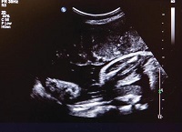
Hydronephrosis may be seen on a routine ultrasound. Follow-up ultrasounds are needed to watch the hydronephrosis, whether it is getting more swollen or is getting better. With a bladder outlet obstruction, doctors often see enlargement of the renal pelvis, a large bladder and both the right and left ureters are swollen with urine that has collected in them also.
The cases to be most concerned about include obstruction on both sides that happens early in the pregnancy with oligohydramnios (lack of amniotic fluid). Amniotic fluid volume is the most important sign of fetal well-being. Another finding that causes concern is an extremely enlarged bladder.
Your obstetrician will likely refer you to a specialist that handles high-risk pregnancies. These doctors are called perinatologists. The perinatologist will perform a targeted ultrasound to confirm the diagnosis and look at the rest of the baby's body for other problems. During this exam, the doctor will look at the overall appearance of each kidney; the amount of swelling in each area of the urinary tract; whether one or both kidneys are affected; overall fetal growth; gender; amniotic fluid index (the amount of amniotic fluid); bladder size; thickness, and emptying; whether there are other birth defects; and presence of fluid in other areas of the body (non-immune hydrops fetalis).
An amniocentesis is a procedure that allows doctors to look at the baby's chromosomes. Your doctor may recommend this procedure because there is a 9 to 12 percent chance of babies with urologic problems having a chromosomal disorder. The perinatologist will discuss the risks and benefits of this procedure with you before it is done.
A fetal echocardiogram may also be recommended to rule out a cardiac defect. This is an ultrasound of the heart's general anatomy, blood flow, vessels and valves. This test would be done by a pediatric heart specialist (cardiologist).
How does bladder outlet obstruction affect my baby?
The urethra is a tube through which urine passes from the bladder and exits the body. In males the most common cause of bladder outlet obstruction is posterior urethral valves (the valves at the opening of the urethra are not positioned correctly). In females the most common cause is urethral atresia (blocked or absent urethra). There is a broad range of severity for bladder outlet obstruction. At one end of the range is normal amniotic fluid volume with functional kidneys. Babies on the other end of the range may have no amniotic fluid, a severely swelled bladder and problems with the kidneys, lungs and other organs.
The problems your baby has will depend on how bad the symptoms are and how far along the pregnancy is when the problems happen. The worse the symptoms are and the sooner they are seen, the worse the outcome tends to be for the baby.
Ultrasound exams are very important to follow how the urinary tract appears and whether this disorder is getting better or worse. In some cases. the problem will improve on its own. In other cases, the problem will get worse. Treatment during the pregnancy, prognosis, as well as plan of care for the baby after delivery will depend on what happens as the pregnancy progresses.
How does bladder outlet obstruction affect my pregnancy?
Hydronephrosis may be seen with a routine ultrasound. Your obstetrician will likely refer you to a specialist who handles high-risk pregnancies for a targeted ultrasound that can examine your unborn baby in more detail. These specialists are called perinatologists. The two most important factors in determining outcome for your baby are the amount of amniotic fluid surrounding the baby and the health (whether they can do their "job") of the kidneys. Each of these factors can change for better or worse as the pregnancy progresses. The plan of care for babies with hydronephrosis will include serial ultrasounds to check on how the baby's kidneys are working. The amount of amniotic fluid will be checked because it is the best clue as to how well the kidneys are working. In most cases, the pregnancy will progress normally.
In a small percentage of cases, the symptoms will worsen. In some cases, the doctor may recommend a procedure be performed on the unborn baby to help kidney function and lung development. If this becomes a treatment option, the procedure's risks and benefits will be discussed in detail with you.
In other cases, the symptoms occur so early in the pregnancy or have become so bad that there is no way to treat it either before or after delivery. If the baby's condition is so bad that he or she cannot survive, two options will be discussed with you. One option is ending the pregnancy early because we know the baby cannot survive and there is no treatment to fix the problem. The other option is continuing the pregnancy with palliative care as the plan of treatment for the baby when he or she is born. The goal of palliative care is support and comfort for the baby and family, and assistance in memory making, bonding and working through the grieving process.
How is bladder outlet obstruction treated?
If bladder outlet obstruction is suspected, a detailed ultrasound exam should be done to look for other birth defects. An amniocentesis may be recommended based on these findings to rule out a genetic disorder. As many as 9 to 12 percent of babies with bladder outlet obstruction may have a genetic disorder.
Bladder outlet obstruction has a wide range of symptoms. In mild cases, there are few changes in their bladder and ureters, no abnormal changes to the kidneys and the amount of amniotic fluid stays within the normal range. In severe cases, there is a lack of amniotic fluid (oligohydramnios), a very swelled bladder and ureters, and abnormal changes to the kidneys.
Because of this wide range of severity, treatment before and after birth will vary also. In mild cases, the place, timing and way the baby is delivered may not be changed. These babies will need follow-up care with a urologist (specialist who treats the urinary tract) and/or nephrologist (specialist who treats disorders of the kidneys) soon after birth. In severe cases, aggressive treatment may not be an option because of the damage to the kidneys and the lack of lung development due to no amniotic fluid. For these babies, palliative care may be the best treatment because the organ damage is too severe for the baby to survive. The goal of treatment will be comfort and helping parents through the bonding and grieving process.
Some hospitals have performed experimental surgery on unborn babies with bladder outlet obstruction to drain the urine out of the kidney(s) or bladder. During this surgery, done on the unborn baby, a tube called a vesicoamniotic shunt is placed to drain the excess urine out of the kidney or bladder and into the amniotic sac that surrounds the baby. In babies with oligohydramnios that has developed over time and after 16 to 24 weeks, early diagnoses and prompt surgery is the best way to help make certain that the lungs develop and mature; and by preventing the back flow of urine this procedure can help preserve kidney function (prevent damage). However, babies that develop oligohydramnios at or before 16 to 24 weeks are not candidates for surgery because it will not help their lung development. These baby's lungs have been severely affected and will not respond to this treatment.
Tests will be done on the urine in the baby's bladder to test how well the kidneys are working before this surgery is considered. The urine is obtained in a procedure similar to an amniocentesis. The perinatologist will discuss the risks and benefits of the procedure before it is done. The urine retrieval is repeated more than once to get "new" urine as opposed to "old" urine that has been sitting in the bladder for some time.
The fetal surgery involves placing a tube in the bladder that will drain the urine out of the baby's body and into the amniotic sac, which holds the baby, placenta and amniotic fluid. Often, more than one tube must be placed because the tubes can become clogged or with the movement of the baby can be moved out of place so that they are no longer draining the urnine. The goal of this surgery is to increase the amount of amniotic fluid, which will help lung development and to maintain kidney function by preventing backflow of urine.
What about after surgery?
After delivery the care provided will depend on the baby's condition. Doctors will measure the urine output and perform a detailed physical exam and blood test that helps gauge how well the kidneys are working. An ultrasound exam of the kidneys and bladder will be done early if hydronephrosis was severe before birth. Voiding cystorurethrography (VCUG) is an X-ray exam that watches urine production and emptying of the bladder. A renal scan may be done to examine for backflow of urine.
One priority soon after birth is finding the diagnosis, which helps determine the treatment plan. The most important goal of treatment in a newborn is making sure the bladder is draining. There are surgeries to help with this, and they are especially important when urinary catheterization (passing a tube through the urethra and into the bladder) is difficult or impossible. This is often the case with PUV.
Posterior urethral valves may at first be treated with endoscopy. Endoscopic treatment uses a scope-type instrument to look at and treat PUV. Treatment is ablation or cautery (removal of tissue) of the valves. Endoscopic treatment is the best treatment but some newborns may have to wait for this procedure until they have grown.
When endoscopic treatment is not an option, a connection between the bladder and lower abdominal wall can be created to provide bladder drainage. This is called a vesicostomy. This surgery is performed to create an external opening for the bladder between the lower abdomen and the belly button. Another option is making an external opening for the ureters. This is called ureterostomies. Any surgery has risks, and the risks and benefits of whatever procedure is determined to be the best for your baby will be discussed with you before the procedure is done.
There also are other treatments that may be part of the baby's care before or after birth. If these are felt to be a treatment that would help your baby the doctor will discuss this with you.
What is my baby's long-term prognosis?
Long-term prognosis for the baby who is diagnosed with bladder outlet obstruction before birth and who has adequate lung development, is improving every day. Lung development, which is measured before birth by the amount of amniotic fluid, is the single most important factor in determining the baby's chance for survival. The death rate for infants diagnosed before birth is 3 percent or less. With early diagnosis, prompt treatment and improved surgery methods for obstruction, the survival rate and quality of life has improved greatly in recent years.
However, there may be some long-term problems for these infants. The most common issues are with bladder function and urinary incontinence, urinary tract infections and kidney damage. Some of these children will have urinary reflux (backflow of urine). These issues can be treated with medication, voiding techniques, intermittent catheterization (passing a tube through the urethra and into the bladder), and/or surgery. Some patients with PUV will eventually require dialysis and a kidney transplant.
Managing bladder function will help maintain kidney function. Medications and voiding techniques will be a lifelong means to control symptoms, but they do not cure the problem. Long-term follow-up care to monitor kidney function and treat issues is of the highest importance for these patients.
Get a second opinion
(414) 240-1831
Research and outcomes
Our outcomes reports help families and partner providers make the most informed healthcare decisions. Learn more about our surgical outcomes and current research studies.
Contact us
For additional information on the Fetal Concerns Center at Children's Wisconsin, please call:
(414) 337-4776
Fax: (414) 337-1884
Note: These phone numbers should not be used for urgent medical concerns. Please contact your physician directly if your situation requires immediate attention, or dial 911 if it is an emergency.

